UDK: 617.7-007.681-073.756.8
Petrushevska A.¹,2, Trpevska Shekerinov N.¹,2, Nikolovski A.¹, Bogdanova I.¹,2.
¹University Clinic of Eye Diseases Skopje, Republic of North Macedonia
2Medical Faculty, „Ss. Cyril and Methodius” University Skopje, Republic of North Macedonia
Abstract
Angle recession glaucoma is classified as a type of traumatic secondary open-angle glaucoma. Angular resection is strongly associated with traumatic hyphema. As many as 60% of nonpenetrating or consecutive ocular trauma will develop some degree of angle resection. The most commonly associated conditions are sports injuries (boxing, paintball), traffic accidents (impact from an airbag, other ocular trauma), physical attacks, falls from a height.
We present a case of a 27-years-old patient injured in a traffic accident in whom we diagnosed secondary glaucoma with a recession of the iridocorneal angle after blunt trauma to the eyeball.
Regression of symptoms and normalization of the condition of the left eye were monitored for one week. At the first control, the visual acuity has improved significantly. When measuring the eye’s pressure of the left eye, an increase in intraocular pressure with a value of 27mmHg was observed. Asymmetry of different sectors of the angle was determined gonioscopically. Subsequently, anterior-segment optical coherence tomography was performed once again, which confirmed the presumed diagnosis.
Key Words: angle resection, glaucoma, ocular trauma, anterior-segment optical coherence tomography.
Introduction
Angle recession glaucoma (ARG) is classified as a type of traumatic secondary open-angle glaucoma. Traumatic glaucoma refer to a heterogeneous group of post-traumatic ocular disorders that, by a different mechanism, lead to an abnormal elevation of intraocular pressure and have an increased risk of developing optic neuropathy (1).
It has been reported that up to 60% of non-penetrating or consecutive ocular trauma will develop some degree of angle resection. The most commonly associated conditions are sports injuries (boxing, paintball), traffic accidents (impact from an air bag, other ocular trauma), physical attacks, falls from a height (1).
Angular resection is strongly associated with traumatic hyphema. As many as 60% of nonpenetrating or consecutive ocular trauma will develop some degree of angle resection. The most commonly associated conditions are sports injuries (boxing, paintball), traffic accidents (impact from an airbag, other ocular trauma), physical attacks, falls from a height (2,3).
ARG was first described by Collins in 1892. The association between ocular trauma and unilateral glaucoma was described by D’Ombrain in 1949 (1).
Case Report
In this paper, we describe a case of a 27-years-old patient in whom we diagnosed secondary glaucoma with recession of iridocorneal angle after blunt trauma to the eyeball. Namely, the patient was injured in a traffic accident.
During inspection, a periocular hematoma was observed on the left eye. The best-corrected visual acuity according to Snellen optotype was 0.16. Eye pressure was digital/ normal.
The motility of the left eye was normal in all directions, without the appearance of double images.
During examination with a biomicroscope, diffuse edema of the cornea, folds of Descemet, slightly dilated pupil temporally and the presence of hyphema in the anterior chamber of the eye were observed. The posterior segment was without ability to trace details. Echography was done, without the presence of pathological content in the vitreous.
The patient was hospitalized, placed on resorptive, antibiotic therapy. Regression of symptoms and normalization of the condition of the left eye were monitored for one week, after which the patient was sent for home treatment.
At the first control, the visual acuity has improved significantly, and at the time the best corrected visual acuity was 1.0. When measuring the eye pressure of the left eye, an increase in intraocular pressure was observed with a value of 27mmHg NCT (pachymetry: 489). Biomicroscopically, the cornea was neat, transparent, anterior chamber was without side contents, pupil slightly mydriatic, temporally at 4h irregular (synechiae), lens neat. Next, gonioscopy with a Goldman lens was approached. Asymmetry of different sectors of the angle was determined gonioscopically. A large area of the ciliary body was visible especially with retrograde depression of the iris (indentation) including the ciliary processes and the circular ciliary muscle. Characteristic deepening and clefting of the angle were noted, which were expected to become more prominent with time due to atrophy and fibrosis of the ciliary muscle.
Subsequently, anterior-segment optical coherence tomography was performed once again, which confirmed the presumed diagnosis.
The patient was placed on anti-glaucomatous therapy (beta-blocker), with which the eye pressure normalized, and it continued to be normal during subsequent control examinations.
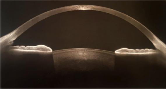
AS-OCT
Discussion
Angular recession is an important clinical sign of previous contusion injury. It is not responsible for glaucoma by itself. Secondary angle-recession glaucoma occurs due to more subtle changes in the trabecular meshwork.
The pathohistological mechanisms that lead to an increase in intraocular pressure are multifactorial. Five key processes are described:
First, blunt force at the time of eyeball injury causes the pupil to be pushed laterally and posteriorly toward the iris and angle. These hydrodynamic forces deepen the iridocorneal angle and increase the diameter of the corneoscleral limbal ring.
A split is caused between the longitudinal and connecting circular and oblique muscles. The tear is usually peripheral to the ciliary body circulus arteriosus and may tear the branches – the anterior and posterior ciliary arteries, the end result of which is bleeding into the anterior chamber. An eye that physiologically has a wider iridocorneal angle has a greater tendency to develop ARG than an eye that has a normal or narrow angle.
Muscles can atrophy years later after the trauma. Degenerative changes resulting in fibrosis and obliteration of the intertrabecular space and Schlemm’s canal have been observed on biomicroscopic examinations. Some studies indicate that the formation of a hyaline membrane that covers the angle and is centrally connected to Descemet’s membrane, may cause posterior expansion of this membrane (4,5,6).
Diagnosing angle recession is not difficult at all. As the surface of the iris is traced to its root, widening of the angle is noted. The rupture occurs in the ciliary body, between the plane of the circular and longitudinal muscle fibers. The iris and circular muscle fibers are seen posteriorly. Longitudinal muscle fibers remain attached to the scleral spur. The gap between the two is clear, and bending of the beam of the cut lamp in the region of the gap is also present. Angle recession differs from cyclodialysis in that the ciliary body detaches from the scleral spur exposing the surface of the sclera in the angle (7).
Ocular trauma is a significant cause of blindness worldwide, especially if associated with glaucoma. Direct damage from blunt or penetrating trauma, hemorrhage, inflammation, lens-related problems, trauma-related orbital and cerebrovascular pathologies, and chemical injuries can increase intraocular pressure, and lead to traumatic glaucoma. Loss of vision can occur due to eye trauma itself or due to its complications. Glaucoma can develop over time due to significant ocular hypertension, which is a major complication of an eye that has had trauma. According to Girkin and colleagues, blunt trauma is more likely to cause glaucoma than penetrating trauma. Cession of the angle of the anterior chamber and its association with monocular chronic simple glaucoma has not been fully appreciated. Ocular hypertension can occur in the short or long term after trauma (7,8).
In clinical practice, assessment of the iridocorneal angle is done using traditional or high-tech methods. Gonioscopy is the traditional method, where a gonio lens are used to examine the angular structures in detail. In addition to being an inexpensive method, gonioscopy allows the evaluation of pigmentation and provides a dynamic assessment of the structures of the indentation angle. However, it is a method that relies heavily on the experience of the operator and the patient’s cooperation, and it requires local anesthesia and careful disinfection to avoid infection. By revolutionizing ophthalmic practice, high-tech methods of diagnosis are used today, such as anterior segment optical coherence tomography (AS-OCT) (9).
Managing ARGs can be challenging. The initial treatment is medical therapy, eventually followed by surgical intervention. The treatment of this secondary glaucoma is in many ways similar to that of primary open-angle glaucoma, although miotic drugs and argon laser trabeculoplasty are controversial therapies for this condition. The only precaution is to avoid the use of prostaglandin analogues in the immediate post-traumatic period, due to their possible pro-inflammatory effect. Surgical treatment is necessary when post-traumatic glaucoma is resistant to medical therapy. In filtration surgery, mitomycin-C is recommended to prevent filtration fibrosis, which is common in eyes with prior trauma (10,11,12).
Conclusion
Following ocular trauma, it is not uncommon for many patients to develop some form of secondary glaucoma. Severe blunt ocular trauma (an ocular contusion) can lead to a glaucomatous state by producing a hyphema, angle recession and inflammation. Despite the gonioscopy in the diagnosis of ARG, AS-OCT plays a significant role as a non-invasive method. Screening for ocular hypertonia must be regular and systematic after ocular trauma involving lesion of the iridocorneal angle.
References:
- De Leon-Ortega JE, Girkin CA. Ocular trauma-related glaucoma. Ophthalmol Clin North Am. 2002 Jun;15(2):215-23. doi: 10.1016/s0896-1549(02)00011-1. PMID: 12229238.
- Ng DS, Ching RH, Chan CW. Angle-recession glaucoma: long-term clinical outcomes over a 10-year period in traumatic microhyphema. Int Ophthalmol. 2015 Feb;35(1):107-13.
- Bansal S, Gunasekeran DV, Ang B, Lee J, Khandelwal R, Sullivan P, Agrawal R. Controversies in the pathophysiology and management of hyphema. Surv Ophthalmol. 2016 May-Jun;61(3):297-308.
- Razeghinejad R, Lin MM, Lee D, Katz LJ, Myers JS. Pathophysiology and management of glaucoma and ocular hypertension related to trauma. Surv Ophthalmol. 2020 Sep-Oct;65(5):530-547.
- Pujari A, Selvan H, Behera AK, Gagrani M, Kapoor S, Dada T. The Probable Mechanism of Traumatic Angle Recession and Cyclodialysis. J Glaucoma. 2020 Jan;29(1):67-70.
- Bansal S, Gunasekeran DV, Ang B, Lee J, Khandelwal R, Sullivan P, Agrawal R. Controversies in the pathophysiology and management of hyphema. Surv Ophthalmol. 2016 May-Jun;61(3):297-308.
- Malik SR, Choudhry S, Singh G. Traumatic recession of the angle of anterior chamber. Indian J Ophthalmol. 1973 Jun;21(2):68-72.
- Iannucci, V.; Manni, P.; Alisi, L.; Mecarelli, G.; Lambiase, A.; Bruscolini, A. Bilateral Angle Recession and Chronic Post-Traumatic Glaucoma: A Review of the Literature and a Case Report. Life 2023, 13, 1814.
- European Glaucoma Society. Terminology and Guidelines for Glaucoma, 5th Edition. Br. J. Ophthalmol. 2021, 105 (Suppl. S1), 1–169.
- AlObaida I, Aljasim LA. Selective laser trabeculoplasty in patients with angle recession glaucoma: A small case series. Am J Ophthalmol Case Rep. 2020 Jul 29;19:100835.
- Fingeret M, Mathews TA, Fodera FA. Angle recession. Optom Clin. 1993;3(2):41-8.
- Bai, H.Q.; Yao, L.; Wang, D.B.; Jin, R.; Wang, Y.X. Causes and Treatments of Traumatic Secondary Glaucoma. Eur. J. Ophthalmol. 2009, 19, 201–206.