UDK: 615.849.1.015.3
Chabukovska Radulovska J1,2
1 University Clinic for Surgical Disease “St. Naum Ohridski“, Skopje,
2 Faculty of Medical Sciences, “Goce Delchev” University, Shtip.
High levels of exposure to ionizing radiation are known to increase the risk of cancer.
The increasing number of computed tomography exams for diagnostic purposes and interventional radiological procedures became a serious source of radiation dose which continues to be one of the hottest topics in radiology, as government bodies and public health concerns push healthcare teams to find ways how to reduce the dose.
For this purpose, the World Health Organization, the IAEA and many other international organizations have prepared certain recommendations for the use of radiation sources, compliance with legal regulations, measurement of doses and strategies for their reduction, without poverty to patients who are the focus of attention (Figure1).

Figure1.
Following the existing European Directive (2013/59/EURATOM) and the increased focus on patients’ safety, the international guidelines and regulations, indicate the necessary need for Dose Monitoring and Management Systems (DMMS), which have already been introduced in the radiological departments for medical radiological diagnostics in several countries in Europe and the world.
In doing so, it is necessary to use Systems for measuring radiation doses and automatic reporting.
There are some limitations in dose monitoring and in accordance with the International: Integrating the Healthcare Enterprise (IHE): “It is of great importance to know the technical limitations, as well as the limitations that make it difficult to monitor the dose in practice, they can explain the reasons why the monitored values do not give the exact dose delivered to the patient. Accordingly to this it is important to understand that by calculating the dose using phantoms and calculating with formulas, it gives the calculated estimated and not the real measured dose. The provide values in that case are only calculated estimates, not “measurements”.
For computed tomography, “CTDI” is the dose estimated on a standard plastic phantom, not on a human tissue. Therefore, the dose should not be represented as the dose received by the patient. For planar or projection images, the recorded values may be exposure, skin dose, or some other value that may not be the patient’s body or organ dose. It is inaccurate to aggregate the estimates of doses which are received from different body parts into a single cumulative value. The concern and attention to monitoring radiation dose estimates is evident in official documents such as: European Directive Euroatom 97/43 and ACR.
In 1981, computed tomography dose index (CTDI) defined as a commonly used index of radiation exposure in X-ray computed tomography (CT). The CTDI unit presented in (Gy), can be used together with the patient’s size to estimate the absorbed dose. CTDI and absorbed dose may differ by more than a factor of two for children which is a small patient.
CTDI vol. is based on measurements obtained during a 16cm or 32cm phantom scan. Basically, it represents the output of the scanner. DLP is derived from CTDI vol. but includes a scan length component. Both work as reasonable values for the absorbed dose, but do not represent the patient’s actual dose. If the CTDI vol. and/or DLP is twice as high as it could be, then the doses the patient receives will be about twice as high as they could be.
HOW TO CALCULATE THE DOSE?
There are several ways to calculate radiation dose measurements and several different units of measurement. This area is still developing as there is no standardization.
But radiation dose constantly changes during the time.
However, there are some methods used to collect data by using the dose information that is captured automatically by the imaging system and by using multipliers to calculate the estimated exposure to a given bad dose.
With the development of the new imaging technology, dose calculation values change and it becomes hard to explain to the patients or physicians why dose parameters for the same type of exam and the same patient change between scans that are years apart.
An important problem in medical radiological diagnostics is the lack of established national guidelines for standard doses (DRLs), so that dose levels can vary significantly.
There is a wide range of variation in protocols at different centers on CT, and within the same hospital, resulting in the same person having a brain CT scan at five different centers receiving five different dose values.
WHY DO WE NEED DOSE MEASUREMENT AND MANAGEMENT SYSTEM?
Radiation dose monitoring in radio diagnostic examinations using X-ray technique is mandatory, to enable reduction of the patient’s exposure to radiation, which is of particular importance in Computed Tomography and interventional radiological procedures.
It is necessary to use Systems for measuring radiation doses and automatic reporting remote Quality Control systems that make the data available to all personnel.
For the purposes of quality control (QC) and quality assurance (QA) in modern radiology, systematic monitoring and analysis of dose-related data from radiological examinations is basic.
Radiation dose monitoring software automatically collects stores and analyzes information about patients’ radiation exposure from medical radiology procedures involving ionizing radiation. Dose management solution can automatically monitor, evaluate and optimize the radiation dose that patients receive.
There are also solutions for centralized technical quality monitoring in digital screening programs, such as e.g., breast cancer. Many systems for measuring and managing radiation doses have such MAMMO solutions in their software.
Information about the dose can be collected directly from the imaging device through a picture archiving and communication system (PACS).
The software uses digital imaging and communications in medicine from (DICOM) -standard data sources.
Obtained data on the radiation dose index can help in the optimization of radiation doses, in order to find the lowest reasonable radiation dose with good radiological image quality. (ALARA), Figure 2.
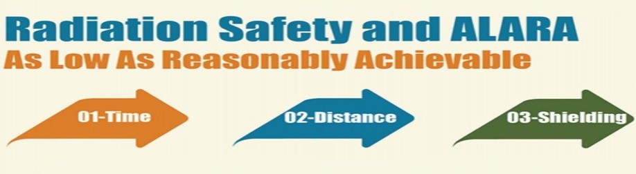
Figure 2.
Dose monitoring systems as a quality management tool enable us the following:
- Real-time performance evaluation and multi-parameter monitoring.
- The procedure can be compared to doses delivered by other procedures with the same
study description or for the same anatomical area.
- Quick correction by prompting the change of wrong test parameters.
- Appropriate alerts can be automatically produced, so that improvement strategies can
be quickly decided upon.
- Staff training and protocol optimization.
- Calculate dose from dose descriptors based on patients’ specific body size, such as
specific dose estimation (SSDE) for CT scans, which takes into account difference between the patient’s actual size and the standard size of the reference phantom (Figure3).
Dose report simulation.

Daily trend – number of patients and mean dose at daily level.

Figure 3.
The management of radiation doses simultaneously provides an opportunity for registration, storage and monitoring of radiation dose, comparison of the information to national DRLs (diagnostic reference levels), issuance of Patient Dose Passports, optimizing patient’s dose and improving quality.
Simultaneous storage of data on radiation doses in the iCloud helps certain coordination in the course of work and availability of large amount of data at the same time, saves time, reduces the workload of data collection staff, facilitates monitoring, creates necessary reports and further improves the quality of health care.
Some of the Dose Monitoring and Management System technologies can help facilitate the management of protocols, personnel dose, usage of contrast media and image quality.
The systematic and continuous monitoring and analysis of radiation dose data can decrease radiation exposure in patients who undergo to the multiple imaging procedures. It can help to meet legal and professional requirements in the hospitals, based on the Medical Exposure Directive 97/43.
In 2019, several hospitals in the Republic of North Macedonia installed software for measuring the radiation doses on CT and mammography devices. They provided information on radiation doses in real time and the possibility of analyzing why certain patients received a higher dose for the same type of examination on the same or on a different device or in a different hospital. The information obtained from this type of monitoring should not scare us but challenge us to be better in the future (Figure 4).

Figure 4.
Conclusion
Software solutions for Dose Measurement and Management are guides for optimized quality, for increased efficiency, compliance with legislation, accreditation and certification, and knowledge management.
Dose monitoring can increase risk awareness among members of the medical staff, which experts considered one of the best ways to improve education of professional workers and raising their awareness for the level of safety procedures for the patient and protection from radiation.
In doing so, the key factors for establishing and maintaining a culture of radiation safety in health care should be defined, in order to improve clinical practice.
References
- www.nice.org.uk/guidance/mib127.
- Loose, R.W., Vano, E., Mildenberger, P. et al. Radiation dose management systems—requirements and recommendations for users from the ESR EuroSafe Imaging initiative. Eur Radiol 31, 2106–2114 (2021). https://doi.org/10.1007/s00330-020-07290-x.
- www.itnonline.com/channel/radiation-dose-management&ved=2ahUKEwi-i-GyjY6GAxVoBdsEHcmXBBQQFnoECBUQAQ&usg=AOvVaw0tBKamrsUIFedzTtp37a7M.
- Loose, R. W., Vano, E., Mildenberger, P., Tsapaki, V., Caramella, D., Sjöberg, J., Paulo, G., Torresin, A., Schindera, S., Frija, G., & Damilakis, J. (2021). Radiation dose management systems—Requirements and recommendations for users from the ESR EuroSafe Imaging initiative. European Radiology, 31(4), 2106-2114. https://doi.org/10.1007/s00330-020-07290-x.
- Vano E, Frija G, Stiller W, et al.; European Society of Radiology (ESR). Harmonisation of imaging dosimetry in clinical practice: practical approaches and guidance from the ESR EuroSafe Imaging initiative. Insights Imaging. 2020 Mar 30;11(1):54. doi: 10.1186/s13244-020-00859-6. PMID: 32232684; PMCID: PMC7105556.
- European Society of Radiology (ESR). Summary of the European Directive 2013/59/Euratom: essentials for health professionals in radiology. Insights Imaging. 2015 Aug;6(4):411-7. doi: 10.1007/s13244-015-0410-4. Epub 2015 May 27. PMID: 26014053; PMCID: PMC4519811.
- EuroSafe Imaging Call for Action 2018, http://www.eurosafeimaging.org/about/call-for-action Accessed 13 Jan 2020
- Järvinen, H., Vassileva, J., Samei, E., Wallace, A., Vano, E., & Rehani, M. (2017). Patient dose monitoring and the use of diagnostic reference levels for the optimization of protection in medical imaging: Current status and challenges worldwide. Journal of Medical Imaging, 4(3). https://doi.org/10.1117/1.JMI.4.3.031214.
- Brambilla M, Vassileva J, Kuchcinska A, Rehani MM (2019) Multinational data on cumulative radiation exposure of patients from recurrent radiological procedures: call for action. Eur Radiol 30. 10.1007/s00330-019-06528-7 [PubMed].
- Homayounieh, F., Holmberg, O., Umairi, R. A., et al. Variations in CT Utilization, Protocols, and Radiation Doses in COVID-19 Pneumonia: Results from 28 Countries in the IAEA Study. Radiology. https://doi.org/10.1148/radiol.2020203453.
- Samara, E. T., Fitousi, N., & Bosmans, H. (2022). Quality assurance of dose management systems. Physica Medica, 99, 10-15. https://doi.org/10.1016/j.ejmp.2022.05.002.
- Duong, P., & Little, B. P. (2014). Dose Tracking and Dose Auditing in a Comprehensive Computed Tomography Dose-Reduction Program. Seminars in Ultrasound, CT and MRI, 35(4), 322-330. https://doi.org/10.1053/j.sult.2014.05.004.
- Pyfferoen L, Mulkens TH, Zanca F, De Bondt T, Parizel PM, Casselman JW (2017) Benchmarking adult CT-dose levels to regional and national references using a dose-tracking software: a multicentre experience. Insights Imaging 8:513–521 [PMC free article] [PubMed].
- Loose RW, Vano E, Mildenberger P, et al; European Society of Radiology (ESR). Radiation dose management systems-requirements and recommendations for users from the ESR EuroSafe Imaging initiative. Eur Radiol. 2021 Apr;31(4):2106-2114. doi: 10.1007/s00330-020-07290-x. Epub 2020 Sep 21. PMID: 32959080; PMCID: PMC7979596.
- European Commission, Food and Agriculture Organization of The United Nations, International Atomic Energy Agency, International Labour Organization, OECD Nuclear Energy Agency, Pan American Health Organization, United Nations Environment Programme, World Health Organization, “Radiation protection and safety of radiation sources: International Basic Safety Standards,” in IAEA Safety Standards Series No. GSR Part 3 (IAEA), Vienna (2014). [Google Scholar].
- Visschedijk, M., Hendriks, R., & Nuyts, K. (2005). How to set up and manage quality control and quality assurance. The Quality Assurance Journal, 9(2), 95-107. https://doi.org/10.1002/qaj.325.
- International Commission on Radiological Protection (ICRP), “Radiological protection in medicine, ICRP Publication 105,” Ann. ICRP 37(6) (2007). 10.1016/j.icrp.2008.08.001 [PubMed] [CrossRef] [Google Scholar].
- Visschedijk, M., Hendriks, R., & Nuyts, K. (2005). How to set up and manage quality control and quality assurance. The Quality Assurance Journal, 9(2), 95-107. https://doi.org/10.1002/qaj.325.
- European Commission (EC), “Medical radiation exposure of the European population,” Radiation Protection No. 180 (2015).
- United Nations Scientific Committee on the Effects of Atomic Radiation (UNSCEAR), “UNSCEAR global survey on medical exposure: a user manual,” 2015.http://www.survey.unscear.org/lib/exe/fetch.php?media=unscear_user_manual_version_may2015.pdf.
- International Atomic Energy Agency, “Dosimetry in diagnostic radiology: an international code of practice,” Technical Reports Series No. 457, IAEA, Vienna: 2007.
- Heilmaier C, Zuber N, Berthold C, Kara L, Weishaupt D. Establishing Local Diagnostic Reference Levels in IR Procedures with Dose Management Software. J Vasc Interv Radiol. 2017 Mar; 28(3):429-441. doi: 10.1016/j.jvir.2016.10.006. Epub 2016 Dec 26. PMID: 28034700.