UDK: 616.711.6-009.7
https://www.doi.org/10.55302/MJA2484118v
Veljanovski D1, Risteski F1, Dungevski G1, Jovanoska I1, Prgova B1, Nanceva A1
1Department of Radiology, General City Hospital “8-mi Septemvri”, Skopje, Republic of North Macedonia
Abstract
A condition known as limbus vertebra occurs when the nucleus pulposus extends beneath the ring apophysis through a gap in the vertebral endplate. While involvement of the inferior and posterior margins is less common, it most frequently occurs at the anterosuperior corner of a single vertebral body, usually in the middle lumbar spine. Thenumber of differential diagnoses, such as tumors, infections or fractures, should be considered.
We discuss the case of a 29-years-old man who was sent to our emergency room following a height-related fall. He claimed palpable pain in his wrist, upper leg, chest and lower back, as well as a brief loss of consciousness. Anterior limbus vertebra (LV) was the diagnosis.
Keywords: Interosseous herniation; fracture; limbus vertebra; low back pain.
Introduction
A condition known as limbus vertebra occurs when the nucleus pulposus protrudes below the ring apophysis through a hole in the vertebra’s endplate (1). While involvement of the inferior and posterior margins is less common, it most frequently occurs at the anterosuperior corner of a single vertebral body, usually in the middle lumbar spine (2).
A smooth, triangular piece of bone known as the ring apophysis may separate as a result of the anterior herniation of the nucleus pulposus. After that, this apophysis remains separated from the spinal body. The limbus vertebra is frequently misdiagnosed as a fracture, which results in needless intrusive operations. What distinguishes a limbus vertebra from a fracture on x-rays is the sclerotic edge surrounding the triangular piece. In contrast, sclerotic borders are absent from acute fractures. When a post-traumatic patient presents with back pain, it is a crucial differential diagnosis (3). In this case, we present a case of a 29-years-old male with LV who was misdiagnosed with a vertebral fracture after presenting with low back discomfort after trauma.
Case Presentation
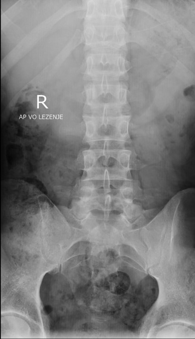
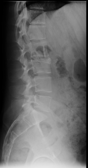
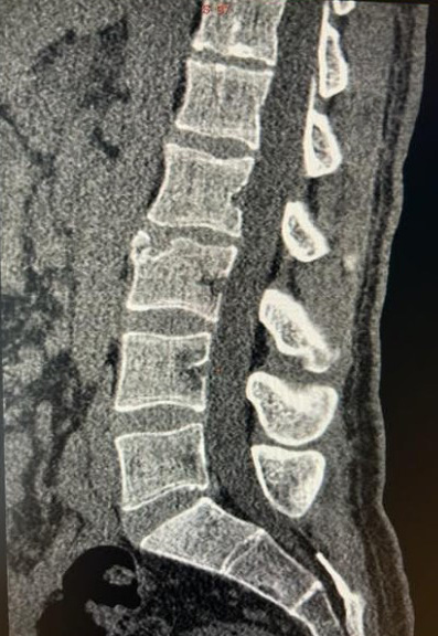
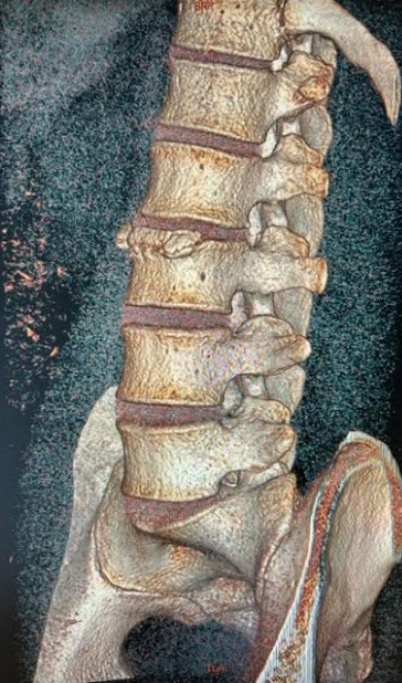
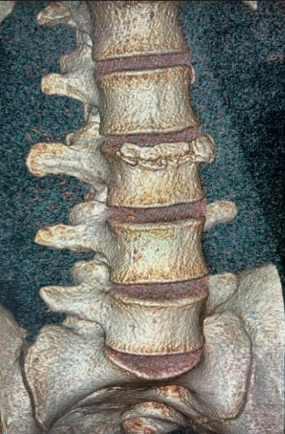
We introduce a 29-years-old man who fell from a height at home and sustained a severe injury in our emergency room. He reported experiencing low back pain, wrist, upper leg and chest palpable discomfort, as well as a brief loss of consciousness. In addition to not being a heavy drinker or smoker, he had no prior pertinent medical history. At the time of the examination, he was hemodynamically stable, able to fully recite the event, aware, and had a soft abdomen that could be palpated. The results of the neurological evaluation were normal. When the lumbar spine was extended and flexed, his range of motion was normal; however, the paravertebral muscles experienced pain and spasms. Mild soreness was felt when the proximal lumbar spine vertebrae were palpated. There were no obvious indications of inflammation. X-rays of the chest, left wrist, thigh and lumbosacral spine were taken; the results showed a fractured left wrist, which was referred for conservative care. A triangular fragment of bone that was corticated and connected to the front and top of the L3 vertebral body was visible on lateral x-rays of the lower back (Figure 1). A second CT scan showed a fractured, triangular piece of hard-edged bone that appeared to have come from a limbus vertebra, as well as rough edge on the cortical material in front of the L3 apophyseal plate with some depression (Figure 2).
A B
Figure 1.Anteroposterior view of the lumbar spine of the patient showing no acute abnormalities (A). Lateral views of the lumbar spine of the patient; white arrows indicate not well separated corticated triangular osseous focus at the anterosuperior aspect of the L3 vertebral body(B).This is the most consistent with a limbus vertebra at L3.
A B C
Figure 2.CT scan showed a cortical marginal irregularity of the L3 apophyseal plates with mild depression with an avulsiontriangular bony focus anteriorly with sclerotic edges compatible with a limbus vertebra(A), 3D volume rendering imaging (B,C).
Discussion
In 1927, Schmorl made the initial identification of the lumbus vertebra and suggested that it was caused by an intrabody disc material herniation that occurred during childhood or adolescence (4,5). Because of its appearance as a triangular bone fragment next to the margin of a vertebral body, plain radiographs frequently misread it as an infection, fracture or tumor. The existence of “disc material” was shown by pathological analyses of these fragments (6). Ghelman and Freiberger used discography in their 1976 work to demonstrate how contrast that was injected into the nucleus pulposus propagated throughout the limbus vertebra. Although reports also indicate the posteroinferior corner and other regional involvement, the anterosuperior edge of a single lumbar vertebral body is the most frequently reported location for the limbus vertebra (7). It is thought that traumas received during childhood and adolescence during the growth of the spine are the cause of limbus vertebrae (8).
The nucleus pulposus may herniate at the border between the ring apophysis and the subsequent vertebral body during this period due to chronic stress, trauma or birth abnormalities, potentially resulting in limbus vertebra (9). In reality, a portion of the ring apophysis ossifies independently after failing to fuse with the rest of the vertebra (10). The disorder is comparable to Schmorl’s nodes, a syndrome in which nuclear material extrudes centrally in the lower thoracic spine and Scheurmann’s illness (11). A simple radiograph may be enough to identify limbus vertebra in adults.
It usually appears as a tiny, triangular, bony mass with a sclerotic surface next to a vertebral body’s edge on x-rays. Particularly for the confirmation of PVL, a condition in which pelvic tissues superimpose with L5 and S1 levels, CT and MRI are regarded as supplementary tests. However, because of its uneven appearance in children and adolescents, this bone segment could be mistaken for an infection or a tumor. Only in cases where imaging appearance is abnormal, additional tests are required for diagnosis. Increased uptake in the vertebral body is seen on a bone scan. By confirming that there is no bone edema, limbus vertebral MRI rules out fractures and suggests a developmental problem (13).
The anterior limbus vertebra (ALV) is frequently discovered by chance in asymptomatic individuals. However, a significant rate of intervertebral disc degeneration (IDD), which is comparable to Scheurmann’s disease, has been observed in teen MRI investigations (13). On the other hand, low back pain was described by Henales et al. in pediatric AVL patients (14). Koyama et al. found a link between ALV and low back discomfort, pointing to risk variables such athletic experience and the COL11A1 genotype (15). Active young athletes are also more susceptible to ALV, according to Acosta et al. (16). ALV appears to develop during childhood and adolescence, according to Baranto et al.’s MRI follow-up on elite athletes, which showed no increase in apophyseal alterations with time (17).
Nerve compression in the posterior limbus vertebra (PVL) might mimic the symptoms of a disc herniation. Lumbus vertebrae frequently don’t require treatment. Non-steroidal anti-inflammatory medicines (NSAIDs), muscle relaxants, and, if required, rehabilitation physical therapy are the usual conservative methods we use to treat symptomatic individuals. Surgical intervention may be required if conservative approaches are not successful. For the limbus fragment to be adequately excised, a total laminectomy is typically necessary (18). Various surgical procedures have also been used by other studies (19, 20). Although Akhaddar et al. suggested removing the mobile fragment while leaving the stable fragment whole, some patients still feel pain following this course of treatment. The results of limbus vertebral surgery vary greatly (21).
Conclusion
Recognizing its characteristic imaging features aids clinicians in making an accurate diagnosis and providing timely treatment. In the differential diagnosis of mechanical lumbar pain, especially in young patients, one should consider the anterosuperior limbus vertebra.
References:
- Ghelman B, Freiberger RH. The limbus vertebra: an anterior disc herniation demonstrated by discography. AJR Am J Roentgenol. 1976;127(5):854-855.
- Mupparapu M, Vuppalapati A, Mozaffari E. Radiographic diagnosis of Limbus vertebra on a lateral cephalometric film: report of a case. DentomaxillofacRadiol. 2002;31(5):328-330.
- Sanal HT, Yilmaz S, Simsek I. Limbus vertebra. Arthritis Rheum. 2012;64(12):4011.
- Schmorl G (1926) Die pathologischeAnatomie der Wirbelsaule. Verhandlungen der DeutchenOrthopadeschenGesellschaft 21: 3-41.
- Schmorl G, Junghanns H (1971) The human Spine in health and disease. (2ndedn), New York, Grune& Stratton.
- Lowrey JJ (1973) Dislocated lumbar vertebral epiphysis in adolescent children. J Neurosurg 38: 232-234.
- Ghelman B, Freiberger RH (1976) The limbus vertebra: an anterior disc herniation demonstrated by discography. AJR Am J Roentgenol 127: 854-855.
- Edelson JG, Nathan H (1988) Stages in the natural history of the vertebral end-plates. Spine (Phila Pa 1976) 13: 21-26.
- Akhaddar A, Belfquih H, Oukabli M, Boucetta M (2011) Posterior ring apophysis separation combined with lumbar disc herniation in adults: a 10-year experience in the surgical management of 87 cases. J Neurosug Spine 14: 475-483.
- Goldman, A, Ghelman B, Doherty J (1990) Posterior limbus vertebrae: a cause of radiating back pain in adolescents and young adults. Skeletal Radiol 19: 501-507.
- Swischuk L, John S, Allbery S (1998) Disk degenerative disease in childhood: Scheuermann’s disease, Schmorl’s nodes, and the limbus vertebra: MRI findings in 12 patients. PediatrRadiolo 28: 334-338.
- Huang PY, Yeh LR, Tzeng WS, Tsai MY, Shih TT, et al. (2012) Imaging features of posterior limbus vertebrae. Clin Imaging 36: 797-802.
- Swischuk L, John S, Allbery S (1998) Disk degenerative disease in childhood: Scheuermann’s disease, Schmorl’s nodes, and the limbus vertebra: MRI findings in 12 patients. PediatrRadiolo 28: 334-338.
- Henales V, Hervas J, Lopez P, Martinez J, Ramos R, et al. (1993) Intervertebral disc herniations (limbus vertebrae) in pediatric patients: report of 15 cases. PediatrRadiol 23: 608-610.
- Koyama K, Nakazato K, Min S, Gushiken K, Hatakeda Y, et al. (2012) COL11A1 gene is associated with limbus vertebra in gymnasts. Int J Sports Med 33: 586-590.
- Acosta V, Pariente E, Lara M, Pini S, Rueda-Gotor J (2016) Limbus Vertebra and Chronic Low Back Pain. J Fam Med 3: 1048-1051.
- Baranto A, Hellstrom M, Cederlund C, Nyman R, Sward L (2009) Back pain and MRI changes in the thoraco-lumbar spine of top athletes in four different sports: A 15-year follow-up study. Knee Surg Sports TraumatolArthrosc 17: 1125-1134.
- Yen Y, Wu F (2014) Clinical Picture: Giant limbus vertebra mimicking a vertebral fracture. Q J Med 107: 937-938.
- Durand D, Huisman T, Carrino J. MR imaging features of common variant spinal anatomy. MagnReson Imaging Clin N Am, 18 (2010), pp. 717-726.
- Scarfo GB, Muzii VF, Mariottini A, Bolognini A, Cartolari R, Posterior retroextramarginal disc hernia (PREMDH): definition, diagnosis, and treatment Surgical neurology1996 46(3):205-11.
- Akhaddar A, Belfquih H, Oukabli M, Boucetta M, Posterior ring apophysis separation combined with lumbar disc herniation in adults: a 10-year experience in the surgical management of 87 cases Journal of neurosurgery Spine2011 14(4):475-83.
